Which figure most likely represents an acoelomate condition? 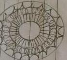 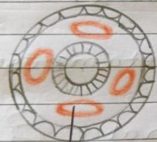 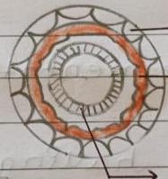 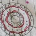
Aimg-5.jpeg

Bimg-6.jpeg

Cimg-7.jpeg

Dimg-3.jpeg

Answer & Solution
Correct answer: A. img-5.jpeg

Principle: an acoelomate has no body cavity between the body wall and the gut; the space is filled with solid tissue.
1. In an acoelomate transverse section, the region between outer ectoderm and inner endoderm appears solid rather than forming a clear cavity.
2. img-5.jpeg shows this solid middle region, matching the condition in platyhelminthes.
3. img-6.jpeg instead shows scattered mesodermal pouches, which is typical of a pseudocoelomate.
4. img-7.jpeg shows a more continuous mesodermal arrangement around a cavity, fitting a true coelomate pattern.
Why the distractors are tempting: img-3.jpeg and img-7.jpeg both show internal spaces, but the presence of a cavity rules out acoelomate organization. img-6.jpeg has incomplete mesodermal lining, but it still represents a pseudocoel, not absence of coelom.
Related questions
A store house of dried plant specimens is called a:In plants, the category corresponding to phylum is called:Chordata is defined by a notochord and a dorsal hollow:Orders Primata and Carnivora are both placed in the class:Felidae and Canidae are included in the order:Convolvulaceae and Solanaceae are included in the order:The genera Panthera and Felis are placed in the family:Solanum, Petunia and Datura are placed in the family: